Meiosis for Middle School Science
- Cate O'Donnell
- May 16, 2025
- 8 min read
Your body is made up of tiny units called cells. Cells are so small that you need a microscope to see them! These cells work together to help you grow, heal, and stay alive. Inside your cells is something very important: your DNA, which carries the instructions that make you who you are. This DNA is organized into chromosomes. Most of your cells have 46 chromosomes, arranged in pairs. These are called body cells. But your body also makes special cells used for reproduction. They are sperm and egg cells, or gametes, and they only have 23 chromosomes. In order to make these cells, your body uses a special process called meiosis.
What Are Chromosomes?
Chromosomes are long, thread-like structures made of DNA and proteins that are found inside the nucleus of most cells. DNA contains the instructions that tell your body how to grow, develop, and function. Because DNA is so long, it is wrapped and folded tightly into compact shapes called chromosomes to fit inside the cell’s nucleus. Humans have 46 chromosomes, arranged in 23 pairs—you get one chromosome in each pair from your mother and one from your father. Each chromosome carries hundreds or even thousands of genes, which are specific sections of DNA that control traits like eye color, height, or how your body digests food.

What Happens During Interphase?
Interphase is the stage of the cell cycle that happens before a cell divides, and it's when the cell prepares for division. This phase takes up most of a cell’s life and is very important because it’s when the cell grows, does its normal jobs, and copies its DNA so it’s ready to split later.
During early interphase, each chromosome in the cell exists as a single, thin strand of DNA. These strands are not tightly coiled yet, so they look more like loose spaghetti inside the nucleus and are not visible under a microscope. Each chromosome is a single copy of DNA and is made up of just one chromatid.
As the cell moves into the middle of interphase, something important happens: the cell copies its DNA. This means that each chromosome now has two identical copies of its DNA, which are called sister chromatids. These chromatids are attached at a place called the centromere, which acts like a belt or button holding them together. At this point, each chromosome looks like an X-shape, with the two arms being the sister chromatids. Even though the DNA has been copied, the sister chromatids stay connected until the cell is ready to divide.
As a cell prepares to divide, it must carefully package its long strands of DNA so they can be moved safely. This process begins throughout interphase, but becomes especially important at the end of interphase. The DNA wraps around special proteins called histones, forming bead-like units called nucleosomes. These nucleosomes coil and fold into thicker and more compact fibers. Just before cell division begins, these fibers become tightly packed into visible chromosomes consisting of the two sister chromatids. This tight coiling helps the chromatids stay organized and makes it easier for the cell to divide the DNA evenly into two new cells.

Prophase I
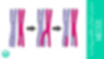
During prophase I of meiosis, several important events prepare the cell for division. First, the chromosomes, which have already duplicated and consist of two sister chromatids, pair up with their matching partner to form what are called homologous pairs. Homologous pairs are two chromosomes—one inherited from the mother and one from the father—that carry the same types of genes in the same order, but the versions of these genes (called alleles) might be different. For example, one chromosome might carry the allele for brown eyes, while the other carries the allele for blue eyes. While paired, the chromosomes exchange sections of DNA in an event called crossing over. This swapping of genetic material creates new combinations of genes, producing recombinant chromosomes, which increases genetic diversity in offspring. Additionally, during prophase I, the nuclear membrane begins to break down, and spindle fibers start to form. These fibers will later help pull the chromosomes apart, ensuring that each new cell receives the correct genetic information.
Metaphase I
During metaphase I of meiosis, the homologous chromosome pairs that formed during prophase I line up along the middle of the cell, called the metaphase plate. Each pair lines up side by side, with one chromosome on one side and its partner on the other. The spindle fibers, which are structures made by the cell to help move chromosomes, attach to the centromeres of each chromosome. This precise arrangement ensures that when the chromosomes are pulled apart in the next phase, each new cell will receive just one chromosome from each homologous pair. This step is crucial for reducing the number of chromosomes by half, which is important for producing sex cells like sperm and eggs.
Anaphase I
During anaphase I of meiosis, the spindle fibers begin to pull the homologous chromosomes apart from each other. Unlike mitosis, where sister chromatids separate, in anaphase I it is the whole chromosomes of each homologous pair that move to opposite ends, or poles, of the cell. The sister chromatids stay attached at their centromeres and move together as one unit. This separation reduces the number of chromosomes in each new cell by half, which is why meiosis is called a reduction division. By the end of anaphase I, each pole of the cell has a mix of maternal and paternal chromosomes, increasing genetic diversity. This step is essential for producing sex cells with the correct number of chromosomes.
Telophase I
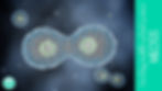
During telophase I of meiosis, the separated homologous chromosomes reach opposite ends, or poles, of the cell. The cell prepares for division by forming a new nuclear membrane around each set of chromosomes, creating two distinct nuclei. However, unlike mitosis, the chromosomes remain duplicated, with sister chromatids still attached. The cell then goes through a process called cytokinesis, where the cytoplasm divides, resulting in two new cells. Each of these cells now has half the number of chromosomes as the original cell, but each chromosome still consists of two sister chromatids. This marks the end of the first meiotic division, and the cells will soon enter meiosis II to separate the sister chromatids.
Interkinesis
Between telophase I and metaphase II in meiosis, the two cells formed after the first division enter a short resting phase called interkinesis. During this time, the cells do not replicate their DNA again. Instead, they prepare for the second division by reorganizing their internal structures. The chromosomes, which are still made up of two sister chromatids, may relax slightly but generally remain condensed. This phase is important because it allows the cells to get ready for the next step in meiosis without duplicating their genetic material again.
Prophase II
During prophase II of meiosis, the two cells formed after meiosis I prepare for the second division. The chromosomes, which are still made up of two sister chromatids, condense again and become visible under a microscope. The nuclear membrane, which may have reformed briefly after telophase I, breaks down again to allow the chromosomes to move freely. Meanwhile, new spindle fibers begin to form from structures called centrosomes at opposite poles of each cell. These spindle fibers will help guide the chromosomes during the next steps. Unlike prophase I, there is no pairing of homologous chromosomes or crossing over because that only happens during the first division.
Metaphase II
During metaphase II of meiosis, the chromosomes, each still made up of two sister chromatids, line up single file along the middle of each of the two cells. This middle line is called the metaphase plate. The spindle fibers, which have formed during prophase II, attach to the centromeres of each sister chromatid. This precise alignment ensures that when the chromatids are pulled apart in the next phase, each new cell will receive one copy of each chromosome. Metaphase II is similar to metaphase in mitosis but occurs in two cells at the same time, preparing the chromatids for separation to form four genetically unique cells.
Anaphase II
During anaphase II of meiosis, the spindle fibers pull the sister chromatids apart at the centromere. Each chromatid, now an individual chromosome, is pulled toward opposite poles of the cell. This separation ensures that each new cell will receive only one copy of each chromosome. Unlike anaphase I, where whole chromosomes were separated, anaphase II divides the sister chromatids, similar to what happens in mitosis. By the end of anaphase II, the cell’s genetic material is divided equally between the two poles, ready for the final stages of meiosis.
Telophase II
During telophase II of meiosis, the separated chromosomes reach the opposite poles of each cell. A new nuclear membrane forms around each set of chromosomes, creating distinct nuclei. The chromosomes begin to uncoil and return to a less condensed form, making them less visible under a microscope. Following this, the cells undergo cytokinesis, where the cytoplasm divides, resulting in a total of four genetically unique haploid cells. Each of these cells contains half the number of chromosomes as the original cell and will develop into sex cells, such as sperm or eggs. This final stage completes meiosis, ensuring genetic diversity in offspring.
Fertilization
When two gametes come together, a process called fertilization occurs. Each gamete, one from the mother (egg) and one from the father (sperm), carries half the number of chromosomes needed for a complete organism. When they join, their genetic material combines to form a single cell called a zygote with a full set of chromosomes. This zygote contains a unique mix of genes from both parents, which means the offspring will have traits inherited from each. Fertilization marks the beginning of a new organism’s development, as the zygote will start dividing through mitosis to grow into a baby. This process ensures that genetic information is passed from one generation to the next.
Chromosomal Disorders
Chromosomal disorders occur when there is a change in the normal number or structure of chromosomes in a person’s cells. Since chromosomes carry the genes that control how our bodies grow and develop, any changes can cause health problems or differences in physical traits. These disorders usually happen because of errors during cell division, especially meiosis. Here are some common examples of chromosomal disorders:
Down Syndrome: This disorder happens when a person has an extra copy of chromosome 21, called trisomy 21. It can cause learning difficulties, distinct facial features, and sometimes health issues like heart problems.
Turner Syndrome: This occurs in females who have only one X chromosome instead of two. It can lead to shorter height, delayed puberty, and fertility challenges.
Klinefelter Syndrome: This affects males who have an extra X chromosome (XXY instead of XY). It can cause taller stature, reduced muscle mass, and sometimes difficulties with speech or learning.
Cri du Chat Syndrome: This disorder is caused by a missing piece of chromosome 5. It is named after the “cat-like” cry babies make, and can cause developmental delays and distinctive facial features.
Understanding chromosomal disorders helps scientists and doctors find better ways to support people living with these conditions.
Meiosis is a special type of cell division that plays a crucial role in sexual reproduction. It reduces the number of chromosomes by half, creating unique sex cells called gametes. This process not only ensures that offspring inherit the correct number of chromosomes from each parent but also increases genetic diversity through the mixing of genes. Without meiosis, life would be much less varied and adaptable. Understanding meiosis helps us appreciate the amazing complexity of life and how traits are passed from one generation to the next.
Want this passage in a printable form? It also comes with my preview, predict, and review page!
Reproduction Flashcards
You can get these flashcards at Teachers Pay Teachers. You can also use them here!